Showing posts with label algae. Show all posts

Friday, May 15, 2015

The Sex Life of Brown Seaweeds


The arrival of spring brings a sudden burst of reproductive growth on the seashore intertidal zone, just as it does in woodlands. The big difference is that on the seashore it's algae - seaweeds - that are switching into reproductive mode, not flowering plants like bluebells. The picture above shows chanelled wrack Pelvetia canaliculata on the shore at Low Newton on the Northumberland coast and ....


.... this is knotted wrack Ascophyllum nodosum. The swollen tips of the channelled wrack and those yellowish egg-shaped objects on the knotted wrack contain the reproductive structures.


Brown seaweeds in the genus Fucus are common in the intertidal zone. Two species are visible here - saw wrack Fucus serratus with a saw-tooth edge to the fronds and bladder wrack F. vesiculosus with smooth frond edges and paired flotation bladders. In spring they make rapid new growth and enter their reproductive phase, producing swollen receptacles at the end of the fronds


The receptacles are covered in large numbers of small swellings called conceptacles, each of which opens via a minute pore called an ostiole. 


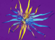
This is a transverse section of the conceptacle of a brown seaweed Fucus sp., viewed under a fluorescence microscope. It has been stained with a fluorescent dye called anilino-naphthalene-sulphonic acid to reveal the detail in its structure. 


This is a section through a receptacle showing two conceptacles developing inside. This is from a female conceptacle. The radiating, elongated filament-like structures are sterile hairs (paraphyses) and the club-shaped structures are oogonia, each of which produces eight eggs (oospheres)....


...... and here is an egg (oosphere) being liberated from an ostiole into the surrounding sea water. Inside the conceptacle some oogonia are still dividing to produce oospheres - you can see the cell walls forming (click on the picture for a larger image).

The clusters of small bright yellow structures that you can see above amongst the rounded oospheres are the antheridia that produce the antherozoids - this conceptacle is hermaphrodite, showing that it came from spiral wrack Fucus spiralis; saw wrack and bladder wrack have conceptacles that are either male or female.

When the conceptacles are mature eggs and vast numbers of swimming male cells (antherozoids) are liberated into the water of the rising tide - most prolifically during spring tides  - and at high water the eggs are fertilised, if they are lucky, and carried away by the falling tide. If they're luckier still the fertilised zygotes attach to a rock and develop into a new seaweed. 


 The pictures above and below show fertile fronds of a Fucus species attached to the harbour wall at St.Peter's marina at the mouth of the river Wear in Sunderland at high tide. That calm water will be seething with countless seaweed eggs and antherozoids, engaged in the frantic business of reproduction. 


You can find more detailed information on the structure and life cycle of seaweeds here  


For soothing movie of seaweeds swaying in the tide click here

Thursday, January 15, 2015

A Golden Wall

Thursday's Guardian Country Diary is about this remarkable 'golden' wall at Hexham, in Northumberland's Tyne valley. It's a retaining wall beside the main Carlisle to Newcastle railway line, that crosses the Tyne just beyond that bridge in the distance and passes this point at head height.


The golden covering is caused by an alga called Trentepohlia aurea.


It spends the drier months of the year as a  powdery deposit on rocks and tree trunks but when it's wet the alga grows into a forest of minute filaments, forming a dense mat on the surface.


You can see the filaments in this close-up, grouped together to form golden cushions that are a few millimetres in diameter. This wall is constantly wet, due to water percolating through from the railway track bed, so conditions are near-perfect for the growth of the alga.


When the tufts coalesce into a golden carpet they provides a very striking back-drop for other plants that are colonising the wall, like this ivy and .....


The liverwort Conocephalum conicum, also known as snakewort. Its surface is divided into small polygons, each with an air pore at its centre, giving it a resemblance to green snakes' skin.


There are other liverworts on the wetter sections of the wall, including this one which I think is Pellia endiviifolia.


The crevices are home to mosses and this little fern with leathery fronds - wall rue Asplenium ruta-muraria.


The alga seems to thrive particularly well on the cement but there are patches of lichen on some of the stones. A close look at this one revealed ....


.... these fungal fruiting bodies, which look like tiny pink toadstools. I think this is a species in the genus Baeomyces.


Another lichen, this time .....


... with fruiting bodies (apothecia)  that look like minute disks of liquorice. A species of Lecidea?


This lichen is a Cladonia species, probably C. fimbriata

Lichens are formed by the symbiotic association between a fungus and an alga and it's very likely that the algal symbiont of some of the lichens on this wall is Trentepohlia


Back now for a closer look at the alga Trentepohlia, this time under the microscope.


Under low magnification (c. x40) with a stereo-microscope you can see the forest of algal filaments that make up those orange cushions, while ....


....... here, under a compound microscope (x100) you can see the cells that make up the filaments, and .....


.... at a higher magnification still (c. x400) you can see the granular, pigmented contents of the cells.

There are animals living in the crevices in this wall, including the fearsome snake-back spider - but that's another story.


Tuesday, June 3, 2014

Snail trail art

Feeding trail left by the abrasive radula (tongue) of a garden snail inside a plastic bucket in our garden. The bucket had been filled with rainwater and developed a lining of slimy algae, which dried on the surface when the water was tipped out - then the snail moved in.